Showing 19–24 of 25 results
-
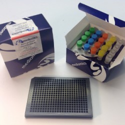
Rabbit IgG FRET-PINCER® Assay Kit, 384-well format
Rabbit IgG FRET-PINCER® Assay Kit, 384-well format
Mediomics’ rabbit immunoglobulin G (rIgG) assay kit can be used to quantitatively detect the concentration of rIgG in biological samples. Product applications include drug and vaccine testing, detection of rIgG antibodies, and determining rIgG concentration. This kit is ideally suited for the rapid, simultaneous measurement of rabbit IgG levels in large number of test samples due to its speed and simplicity. In comparison to the ELISA procedure, the FRET-PINCER® technology provides a simple (one mixing step) and fast (30 minute incubation) assay that is quantitative and reproducible.
-
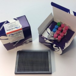
Rabbit IgG TRF-PINCER® Assay Kit, 384-well format
Rabbit IgG TRF-PINCER® Assay Kit, 384-well format
Mediomics’ rabbit immunoglobulin G (rIgG) assay kit can be used to quantitatively detect the concentration of rIgG in biological samples. Product applications include drug and vaccine testing, detection of rIgG antibodies, and determining rIgG concentration. This kit is ideally suited for the rapid, simultaneous measurement of rabbit IgG levels in large number of test samples due to its speed and simplicity. In comparison to the ELISA procedure, the FRET-PINCER® technology provides a simple (one mixing step) and fast (30 minute incubation) assay that is quantitative and reproducible.
-
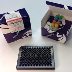
Rabbit IgG TRF-PINCER® Assay Kit, 96-well format
Rabbit IgG TRF-PINCER® Assay Kit, 96-well format
Mediomics’ rabbit immunoglobulin G (rIgG) assay kit can be used to quantitatively detect the concentration of rIgG in biological samples. Product applications include drug and vaccine testing, detection of rIgG antibodies, and determining rIgG concentration. This kit is ideally suited for the rapid, simultaneous measurement of rabbit IgG levels in large number of test samples due to its speed and simplicity. In comparison to the ELISA procedure, the FRET-PINCER® technology provides a simple (one mixing step) and fast (30 minute incubation) assay that is quantitative and reproducible.
-
TR-FRET Bridge-It® L-Methionine (L-Met) Fluorescence Assay Kit, 384-well format
TR-FRET Bridge-It® L-Methionine (L-Met) Fluorescence Assay Kit, 384-well format
The Mediomics Bridge-It® L-methionine fluorescence assay method is based on a combination of well-established fluorescence measurement techniques and a new assay platform design that utilizes DNA-binding proteins as biosensors for their respective small molecule coregulators (ligands). The affinity of the DNA sequence-specific MetJ methionine repressor protein for its unique DNA binding site is greatly increased in the presence of its ligand, S-adenosyl methionine (SAM). For this assay, the MetJ consensus sequence was split into two approximately equal DNA “half-sites” with one half fragment labeled with fluorescein and the other half fragment labeled with Oyster® 645 fluorophore3,4. The relative amount of SAM present in a test sample will influence the amount of DNA-MetJ protein complex formation in the assay. When this complex forms, it brings the fluorescence labeled-DNA half-sites into close proximity and causes a measurable change (increase) in fluorescence signal emission that can be readily measured using a microplate reader (wavelength settings: absorption 485 nm; emission 665 nm).
-
TR-FRET Bridge-It® L-Tryptophan Fluorescence Assay Kit for Plasma and Serum Samples, 384-well format
TR-FRET Bridge-It® L-Tryptophan Fluorescence Assay Kit for Plasma and Serum Samples, 384-well format
The Bridge-It® tryptophan fluorescence assay is based on the activity of tryptophan repressor protein (TrpR), a bacterial DNA-binding protein. TrpR protein binds to its DNA-binding site in tryptophan-dependent fashion.
The central feature of this assay design is the TrpR-dependent association of two fluorochrome-labeled DNA half-fragments (one labeled with fluorescein and the other labeled with Oyster® 645 fluophore). Each fragment contains about one-half of the TrpR protein DNA-binding site. In the presence of L-tryptophan an increase in fluorescence signal can be detected as a result of the tryptophan-dependent association of the labeled DNA half-fragments. Tryptophan is readily detectable using the Bridge-It® tryptophan fluorescence assay in various types of test samples including bacterial growth medium, brain extract, yeast extract, as well as in human serum and urine.
-
TR-FRET Bridge-It® L-Tryptophan Fluorescence Assay Kit, 384-well format
TR-FRET Bridge-It® L-Tryptophan Fluorescence Assay Kit, 384-well format
The Bridge-It® tryptophan fluorescence assay is based on the activity of tryptophan repressor protein (TrpR), a bacterial DNA-binding protein. TrpR protein binds to its DNA-binding site in tryptophan-dependent fashion.
The central feature of this assay design is the TrpR-dependent association of two fluorochrome-labeled DNA half-fragments (one labeled with fluorescein and the other labeled with Oyster® 645 fluophore). Each fragment contains about one-half of the TrpR protein DNA-binding site. In the presence of L-tryptophan an increase in fluorescence signal can be detected as a result of the tryptophan-dependent association of the labeled DNA half-fragments. Tryptophan is readily detectable using the Bridge-It® tryptophan fluorescence assay in various types of test samples including bacterial growth medium, brain extract, yeast extract, as well as in human serum and urine.