Showing all 4 results
-
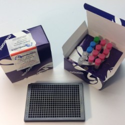
Bridge-It® L-Methionine (L-Met) Fluorescence Assay Kit, 384-well format
Bridge-It® L-Methionine (L-Met) Fluorescence Assay Kit, 384-well format
The Mediomics Bridge-It® L-methionine fluorescence assay method is based on a combination of well-established fluorescence measurement techniques and a new assay platform design that utilizes DNA-binding proteins as biosensors for their respective small molecule coregulators (ligands). The affinity of the DNA sequence-specific MetJ methionine repressor protein for its unique DNA binding site is greatly increased in the presence of its ligand, S-adenosyl methionine (SAM). For this assay, the MetJ consensus sequence was split into two approximately equal DNA “half-sites” with one half fragment labeled with fluorescein and the other half fragment labeled with Oyster® 645 fluorophore3,4. The relative amount of SAM present in a test sample will influence the amount of DNA-MetJ protein complex formation in the assay. When this complex forms, it brings the fluorescence labeled-DNA half-sites into close proximity and causes a measurable change (increase) in fluorescence signal emission that can be readily measured using a microplate reader (wavelength settings: absorption 485 nm; emission 665 nm).
-
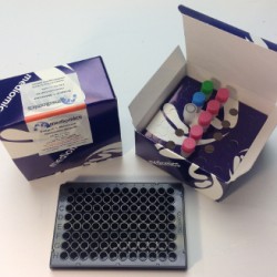
Bridge-It® L-Methionine (L-Met) Fluorescence Assay Kit, 96-well format
Bridge-It® L-Methionine (L-Met) Fluorescence Assay Kit, 96-well format
The Mediomics Bridge-It® L-methionine fluorescence assay method is based on a combination of well-established fluorescence measurement techniques and a new assay platform design that utilizes DNA-binding proteins as biosensors for their respective small molecule coregulators (ligands). The affinity of the DNA sequence-specific MetJ methionine repressor protein for its unique DNA binding site is greatly increased in the presence of its ligand, S-adenosyl methionine (SAM). For this assay, the MetJ consensus sequence was split into two approximately equal DNA “half-sites” with one half fragment labeled with fluorescein and the other half fragment labeled with Oyster® 645 fluorophore3,4. The relative amount of SAM present in a test sample will influence the amount of DNA-MetJ protein complex formation in the assay. When this complex forms, it brings the fluorescence labeled-DNA half-sites into close proximity and causes a measurable change (increase) in fluorescence signal emission that can be readily measured using a microplate reader (wavelength settings: absorption 485 nm; emission 665 nm).
-
TR-FRET Bridge-It® L-Methionine (L-Met) Fluorescence Assay Kit, 384-well format
TR-FRET Bridge-It® L-Methionine (L-Met) Fluorescence Assay Kit, 384-well format
The Mediomics Bridge-It® L-methionine fluorescence assay method is based on a combination of well-established fluorescence measurement techniques and a new assay platform design that utilizes DNA-binding proteins as biosensors for their respective small molecule coregulators (ligands). The affinity of the DNA sequence-specific MetJ methionine repressor protein for its unique DNA binding site is greatly increased in the presence of its ligand, S-adenosyl methionine (SAM). For this assay, the MetJ consensus sequence was split into two approximately equal DNA “half-sites” with one half fragment labeled with fluorescein and the other half fragment labeled with Oyster® 645 fluorophore3,4. The relative amount of SAM present in a test sample will influence the amount of DNA-MetJ protein complex formation in the assay. When this complex forms, it brings the fluorescence labeled-DNA half-sites into close proximity and causes a measurable change (increase) in fluorescence signal emission that can be readily measured using a microplate reader (wavelength settings: absorption 485 nm; emission 665 nm).
-
TR-FRET Bridge-It® L-Methionine (L-Met) Fluorescence Assay Kit, 96-well format
TR-FRET Bridge-It® L-Methionine (L-Met) Fluorescence Assay Kit, 96-well format
The Mediomics Bridge-It® L-methionine fluorescence assay method is based on a combination of well-established fluorescence measurement techniques and a new assay platform design that utilizes DNA-binding proteins as biosensors for their respective small molecule coregulators (ligands). The affinity of the DNA sequence-specific MetJ methionine repressor protein for its unique DNA binding site is greatly increased in the presence of its ligand, S-adenosyl methionine (SAM). For this assay, the MetJ consensus sequence was split into two approximately equal DNA “half-sites” with one half fragment labeled with fluorescein and the other half fragment labeled with Oyster® 645 fluorophore3,4. The relative amount of SAM present in a test sample will influence the amount of DNA-MetJ protein complex formation in the assay. When this complex forms, it brings the fluorescence labeled-DNA half-sites into close proximity and causes a measurable change (increase) in fluorescence signal emission that can be readily measured using a microplate reader (wavelength settings: absorption 485 nm; emission 665 nm).


