TR-FRET
Showing all 7 results
-

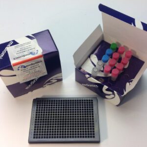
TR-FRET Bridge-It® L-Methionine (L-Met) Fluorescence Assay Kit, 384-well format
$175.00 – $525.00 -
TR-FRET Bridge-It® L-Methionine (L-Met) Fluorescence Assay Kit, 96-well format
$220.00 – $400.00 -
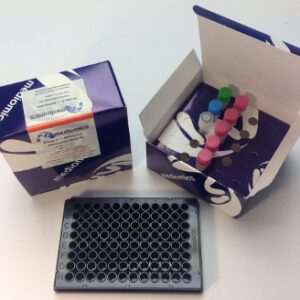
TR-FRET Bridge-It® L-Tryptophan Fluorescence Assay Kit for Plasma and Serum Samples, 384-well format
$175.00 – $525.00 -
TR-FRET Bridge-It® L-Tryptophan Fluorescence Assay Kit, 384-well format
$175.00 – $525.00 -
TR-FRET Bridge-It® L-Tryptophan Fluorescence Assay Kit, 96-well format
$220.00 – $400.00 -
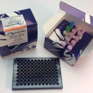
TR-FRET Bridge-It® S-Adenosyl Methionine (SAM) Fluorescence Assay Kit, 384-well format
$175.00 – $525.00 -
TR-FRET Bridge-It® S-Adenosyl Methionine (SAM) Fluorescence Assay Kit, 96-well format
$220.00 – $400.00